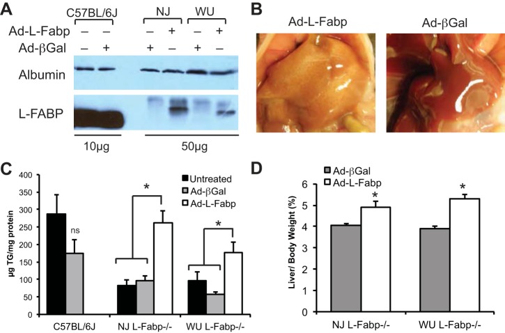
Fig. 2.
Restoration of fasting-induced steatosis by adenoviral L-Fabp. A: expression of L-FABP protein in livers of C57BL/6J mice with or without Ad-βGal (10 μg protein/lane) and in L-Fabp−/− mice injected with Ad-βGal or Ad-L-Fabp (50 μg protein/lane). Exogenous L-Fabp (from Ad-L-Fabp transduction) migrates slower than endogenous L-Fabp (C57BL/6J) because of the addition of a FLAG epitope tag. Expression of albumin is shown as a loading control. B: livers of WU L-Fabp−/− mice injected with either Ad-L-Fabp (left) or Ad-βGal (right) following a 48-h fast. C: hepatic TG in untreated (solid bars), Ad-βGal injected (shaded bars), and Ad-L-Fabp (open bars) injected C57BL/6J and NJ and WU L-Fabp−/− mice following a 48-h fast. D: liver weight in fasted Ad-βGal and Ad-L-Fabp mice expressed as percentage of final body weight. All NJ Fabp−/− mice for this experiment are F1; WU Fabp−/− mice are N8, F9 or N8, F10. Values are means ± SE; no. of mice per group: untreated controls, n = 5–7/genotype, Ad-βGal, n = 3–5/genotype; Ad-L-Fabp, n = 5–6/genotype. *P < 0.05 vs. Ad-βGal and untreated controls.

