Fig. 3.
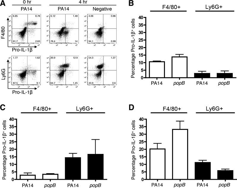
Macrophages and neutrophils temporally recruited to the peritoneum produce pro-IL-1β upon ex vivo infection. C57BL/6 mice were injected with thioglycollate and peritoneal cells were collected by lavage at 0 h (naïve uninjected), 4 h, or 3 days. The harvested cells were infected with WT or popB PA14 at MOI = 0.2 and analyzed 3 h postinfection. Bacteria were excluded from analyses by gating based on scatter controls, and the percentage of pro-IL-1β+F4/80+ and pro-IL-1β+Ly6G+ cells was determined within the populations of peritoneal cells. A: representative dot plot for pro-IL-1β+ cells at 0 or 4 h. B–D: percentage of pro-IL-1β+F4/80+ and pro-IL-1β+Ly6G+ peritoneal cells from 0 h (B), 4 h (C), or 3 days (D) upon ex vivo infection. Data are expressed as means ± SD. Results are representative A, or compiled from independent biological experiments (A–D; n = 3).

