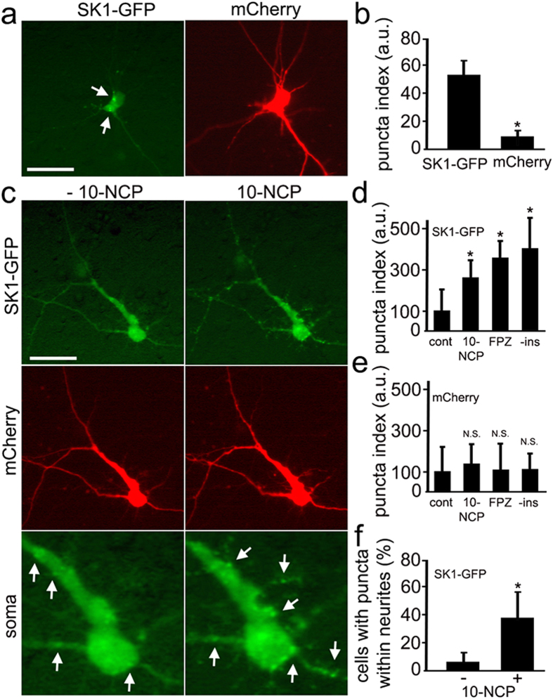
Figure 2. Pharmacological stimulation of autophagy causes the formation of SK1-positive puncta in primary neurons.
(a) In a significant percentage of primary rat cortical neurons, ectopically expressed SK1-GFP exhibits a punctate appearance (left panel), whereas the distribution of a co-transfected marker of morphology mCherry (right panel) is diffuse. Bar, 50 μm. (b) Percentage of neurons with SK1-GFP- and mCherry-positive puncta. *P < 0.01 (t-test). (c) Stimulation of autophagy with 5 μM 10-NCP promotes the formation of SK1-GFP-positive puncta. Live neurons transfected with SK1-GFP and mCherry were observed before (left panel) and after (right panel) a 4-h incubation with 10-NCP. Bar, 50 μm. See bottom panel for higher magnification image of SK1 puncta. Puncta before and after the treatment are depicted with arrows. (d) SK1-GFP-positive puncta were efficiently formed in cortical neurons following treatment with 5 μM 10-NCP or with 5 μM fluphenazine or by incubation in insulin-free medium as reflected by the puncta index. The puncta index was estimated by measuring the standard deviation of SK1-GFP fluorescence in a region corresponding to the neuronal soma and major neurites before and after treatment with 10-NCP (5 μM, 4 h) or with fluphenazine (FPZ; 5 μM, 4 h) or after incubation in insulin-free media (-ins; 4 h). *P < 0.001 (Dunnett’s test). N.S.—non-significant (t-test). (e) The puncta index was estimated by measuring the standard deviation of mCherry fluorescence in the neuronal soma and major neurites. N.S.—non-significant (t-test). (f) Stimulation of autophagy with 10-NCP increases the number of SK1-GFP puncta not only in the soma, but also in neurites. *P < 0.001 (t-test).

