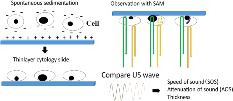
Figure 1. Study design. Free cells of effusion were fixed in 95% ethanol and centrifuged to make precipitates.
Then, the precipitates were washed in distilled water, centrifuged again and poured on a glass slide. The negatively charged cells spontaneously settled on a positively charged slide to form a thin-layer specimen. The cytologic specimen was scanned with US probe to compare the US waves from the surface of the cell and glass slide. The speed of sound (SOS) and the attenuation of sound (AOS) through cells and the thickness of cell were calculated to generate images on screen.

