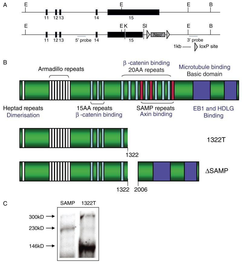
Figure 2.
Construction of ΔSAMP mice. (A) The top panel shows the wild-type allele and the lower panel shows the targeted ΔSAMP allele. Exons are numbered and restriction enzymes marked (B = BamHI; E = EcoR1; K = KpnI; Sl = SalI). (B) A cartoon showing the protein structure of APC and the deleted regions in the 1322T and ΔSAMP mutated proteins. (C) Western blot using anti-Apc antibody on tail fibroblasts derived from heterozygous 1322T and ΔSAMP mice showing the sizes of the mutated Apc protein relative to the full length 300 kD protein.

