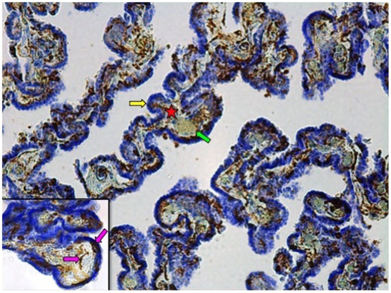
Figure 1.
Iba-1 immunostained choroid plexus with cresyl violet counterstain at 10× magnification. Star denotes connective tissue stroma; yellow arrow indicates epithelial cell layer; green arrow points to blood vessel. Inset: 40× magnification. Pink arrows show Iba1-IR cells associated with the stroma or the epithelial cell layer.

