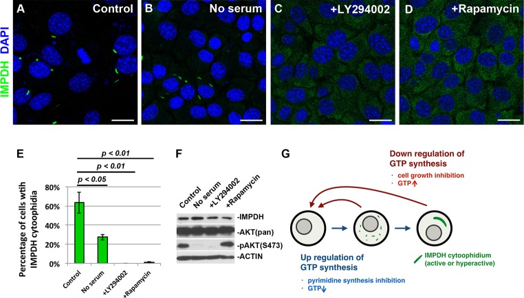
Fig. 3.
IMPDH forms cytoophidia in specific cell types in vitro. (A–D) Immunofluorescence of mouse BNL CL2 cells that were untreated (control), cultured with serum-free medium for 1 day, or treated with 50 μM LY294002 (PI3K inhibitor) or 1 μM rapamycin (mTOR inhibitor) for 6 h before fixation. Scale bars: 20 μm. (E) Mean±s.e.m. percentages of cells with IMPDH cytoophidia after culture under various conditions. P-values were calculated with a Student's t-test. (F) Immunoblotting of BNL CL2 cells cultured under various conditions. AKT(pan), total ATK; pAKT, phosphorylated AKT. (G) A model for the regulation of IMPDH cytoophidium assembly.

