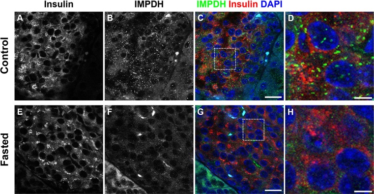
Fig. 4.

IMPDH forms cytoophidia in mouse pancreatic islet cells. (A–C) Immunofluorescence on a normal mouse pancreas section showing abundant immature IMPDH cytoophidia in islet cells. Scale bar: 20 μm. (D) Magnified view of boxed area in C. Scale bar: 5 μm. (E–G) Immunofluorescence on a section of pancreas from a mouse fasted overnight shows that the number of IMPDH cytoophidia in islet cells decreased. Scale bar: 20 μm. (H) Magnified view of boxed area in G. Scale bar: 5 μm.
