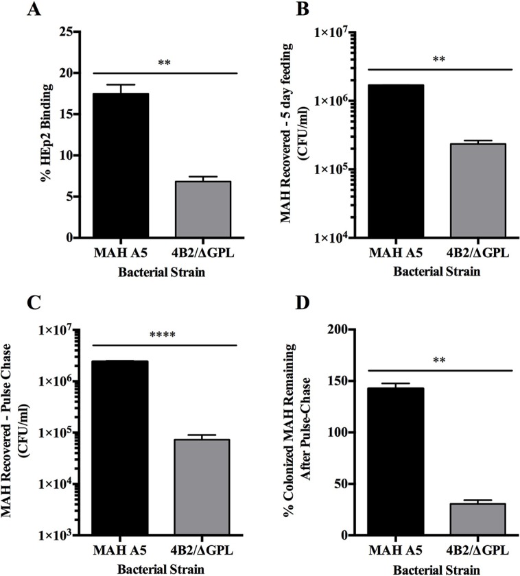
Fig. 6.

Binding of HEp-2 cells and colonization of C. elegans by MAH ΔGPL/4B2 mutant. (A) The MAH 4B2/ΔGPL mutant and the parental strain MAH A5 were used for HEp-2 binding assays. HEp-2 cells were infected at an MOI of 10:1 with each strain and binding was allowed to progress for 1 h at 4°C. Wells were lysed and quantified for percent of bound bacteria to the surface of epithelial cells during assay. Equivalent numbers of C. elegans were seeded onto NGM-FUdR (400 µM) plates containing 108 of each MAH strain and allowed to feed at 25°C for 5 days. Worms were collected, washed with levamisole (25 mM), treated with amikacin (200 µg/ml), and lysed for quantification of intestinal bacteria. (B) Worms were homogenized immediately after MAH feeding to determine intestinal binding ability after feeding. (C) Analysis for colonization using pulse-chase assays were conducted by transferring worms to NGM-FUdR plates seeded with E. coli strain OP50 for 24 h prior to homogenization and quantification. (D) The percent of each MAH strain remaining from the initial 5 days feeding after the pulse-chase was conducted was calculated by (MAH recovered−5 day feeding/MAH recovered−Pulse-Chase)×100. Data represent the mean±s.e.m. of 2 independent experiments each performed in triplicate (**P<0.01, ****P<0.0001 as determined by Student's t-test).
