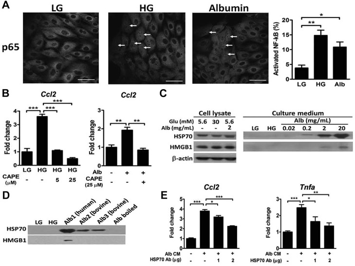
Fig. 3.
Effects of HG and albumin on NF-κB activation and DAMP release in LLC-PK1 cells. (A) Immunofluorescence staining of NF-κB in LLC-PK1 cells after 24 h incubation in medium containing 5.6 mM glucose (LG), 30 mM glucose (HG) or 5.6 mM glucose with 2 mg/ml albumin. The percentage of NF-κB translocation into the nucleus is presented. n=3 in each group. Scale bars: 10 μm. (B) Expression of Ccl2 in LLC-PK1 cells treated with LG, HG or albumin with or without caffeic acid phenethyl ester (CAPE) for 24 h. n=3 in each group. (C) Immunoblot analyses of HSP70 and HMGB1 from the cell lysate and culture medium of LLC-PK1 cells treated with LG, HG and different concentrations of albumin for 24 h. (D) Immunoblot analyses of HSP70 and HMGB1 from the culture medium of LLC-PK1 cells treated with various sources of albumin (2 mg/ml). Alb1, fatty acid free human albumin; Alb2, fatty acid free bovine albumin; Alb3, essential fatty acid free bovine albumin; Alb boiled, Alb3 boiled for 10 min. (E) Expression of Ccl2 and Tnfα in LLC-PK1 cells treated with the conditioned medium (CM) with or without depletion of HSP70 for 8 h. *P<0.05, **P<0.01 and ***P<0.001 by one-way ANOVA followed by Dunnett's test.

