Fig. 4.
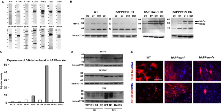
Altered splicing of tau occurs during differentiation of hAPPsw+/+ RGs into astrocytes. (A) Expression of tau antibodies in porcine adult brain reveals the presence of several isoforms of total and phosphorylated tau. (B) Altered splicing of tau is detected using anti-PHF-6 recognizing phosphorylated tau at Thr231 in both hAPPsw+/+ cell lines both prior to and during differentiation. An additional band of 54 kDa is observed and increased expression of 48 kDa is observed in the R4 hAPPsw+/+ line. (C) Levels of expression of the 54 kDa band reveal a significant increase in the R5 hAPPsw+/+ cell line. Adjusted density is calculated by normalization to β-Actin and then adjusted by normalization to R4 hAPPsw+/+ undifferentiated RGs. (D) Western blot confirmed that the 54 kDa band of interest is not EF1-α, SEPTIN7 or VIM, which were predicted proteins following mass spectrometry. This was evident as expression of these proteins were detected in the WT and R1 hAPP+/− RGs. (E) Expression of total tau and pSer199/202 tau varied between the cell lines and revealed that hyperphosphorylated tau was present only in mitotically dividing hAPPsw+/+ cells following a 48-day differentiation into cortical neurons. Magnification: 40×; scale bar: 100 µm.

