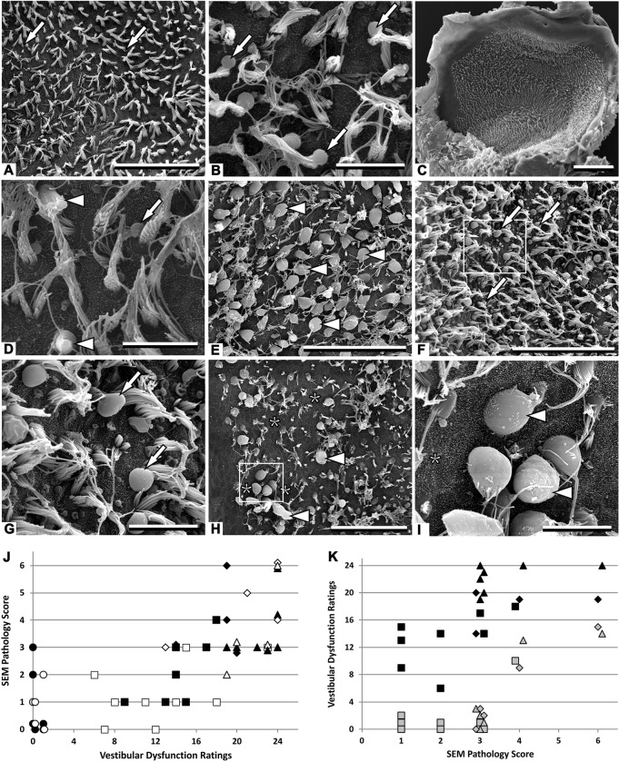
Fig. 2.
Effects of chronic ototoxic exposure (20 mM IDPN in drinking water) and washout on the vestibular sensory epithelium as observed in surface preparations of utricles examined by SEM. (A) Control rat. Only a few small blebs behind stereociliary bundles (arrows) are noted as possible pathological feature in this control sample. (B) Rat exposed for 4 weeks, and with a vestibular dysfunction rating (VDR) of 14. Blebs behind stereociliary bundles (arrows) were the single noticeable pathological feature. (C) Rat exposed for 4 weeks, with a VDR of 23, displaying a control-like appearance at low magnifications. (D) Higher magnification view of the utricle in C revealing only a few pathological features. In addition to blebs (arrow), a modest proportion of stereociliary bundles showed coalescence (arrowheads). (E) Rat exposed for 10 weeks, with a VDR of 24. Note that the majority of the HCs (arrowheads) are extruding into the endolymphatic cavity through the stereociliary bundle. (F) Rat exposed for 6 weeks examined after a washout period of 6 weeks. This animal showed a good recovery: VDR decreased from 24 at the end of the exposure to 3 at the time of histological examination. Blebs (arrows) were abundant and large, but little evidence of HC extrusion or loss was present. (G) Higher magnification view of the boxed area in F. Arrows indicate blebs. (H) Rat exposed for 6 weeks examined after a washout period of 6 weeks. This animal failed to recover well: VDR only varied from 24 at the end of the exposure to 14 at the time of histological examination. Extruding HCs (arrowheads) were abundant and areas containing only supporting cells (asterisks) denoted the loss of HCs. (I) Higher magnification view of the boxed area in H. Arrowheads indicate extruding HCs; asterisk indicates areas containing only supporting cells. (J) Relationship between VDRs and SEM Pathology Scores in control animals (circles) and in animals treated with 20 mM IDPN for 4 (squares), 6 (triangles) or 10 (rhombus) weeks; animals were examined at the end of the exposure (open symbols) or after a washout period (filled symbols). (K) Relationship between SEM Pathology Scores and VDR recovery in animals exposed to 20 mM IDPN and examined after a washout period. Two symbols are included for each animal, showing the VDR at the end of the exposure period (black), and at the end of the washout period (gray). Symbol shape as in G. Scale bars: 50 µm in A,E,F and H; 10 µm in B,D,G and I; 100 µm in C.

