Fig. 6.

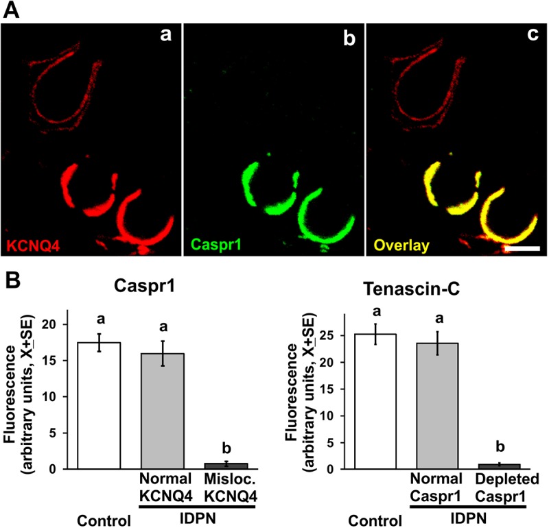
Cell-to-cell basis of calyceal junction dismantlement. (A) A vestibular epithelium from a rat exposed to 10 mM IDPN for 10 weeks. Note the two calyx units showing a normal distribution of KCNQ4 in the inner membrane of the calyx ending (bottom units), and the unit showing abnormal distribution (upper-left unit). CASPR1 immunofluorescence is normal in the first case and highly reduced in the second case (b,c). Scale bar: 5 µm. (B) Left: Quantitative analysis of CASPR1 fluorescence in normal and altered calyx units, as defined by their KCNQ4 distribution, demonstrating that unaffected calices from exposed animals have control-like CASPR1 content. Right: Quantitative analysis of tenascin-C fluorescence in normal and altered calyx units, as defined by their CASPR1 content, demonstrating that unaffected calices from exposed animals have control-like tenascin-C content. In both B and C, n=30 cells from 2-3 animals per group. a,b: groups not sharing a letter are significantly different, P<0.05, Duncan's test after significant ANOVA.
