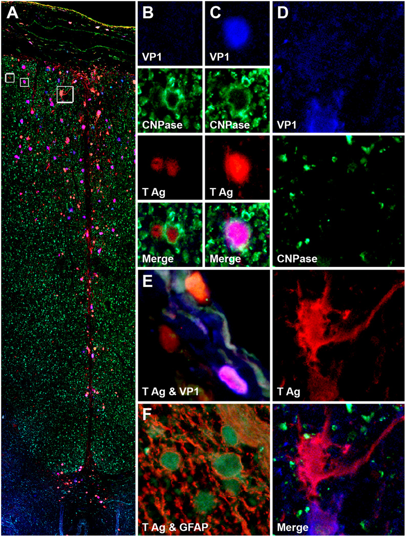
Figure 2.
Large lesion of progressive multifocal leukoencephalopathy in spinal cord. (A-D) Triple immunofluorescence assay for 2',3'-cyclic-nucleotide 3'-phosphodiesterase (CNPase, Alexa Fluor® 488), SV40 T Ag (Alexa Fluor® 568) and VP1 (Alexa Fluor® 350) in monkey #2 shows SV40-infected cells expressing T antigen (T Ag) alone (red), VP1 alone (blue) or both T Ag and VP1 (purple) in the leptomeninges and along the posterior fissure of the spinal cord (A). Magnification, separate channels and merge for the 2 boxes on the left in (A) are shown in (B) and (C). The boxes contain SV40-infected oligodendrocytes (CNP-ase-positive) expressing T Ag alone (B) or T Ag and VP1 (C). (D) Magnification, separate channels and merge for the box on the right in (A) are shown in 4 panels on the right of the montage. This box shows a T Ag-positive SV40-infected cell not stained for CNPase that has an astrocyte phenotype. (E, F) Similarly to other SV40-infected cells, meningeal cells (E) express both T Ag and VP1 separately or simultaneously. A double immunofluorescence assay for T Ag (green) and glial fibrillary acidic protein (GFAP, red) confirms that the majority of SV40-infected cells are astrocytes (F).

