Fig. 1.
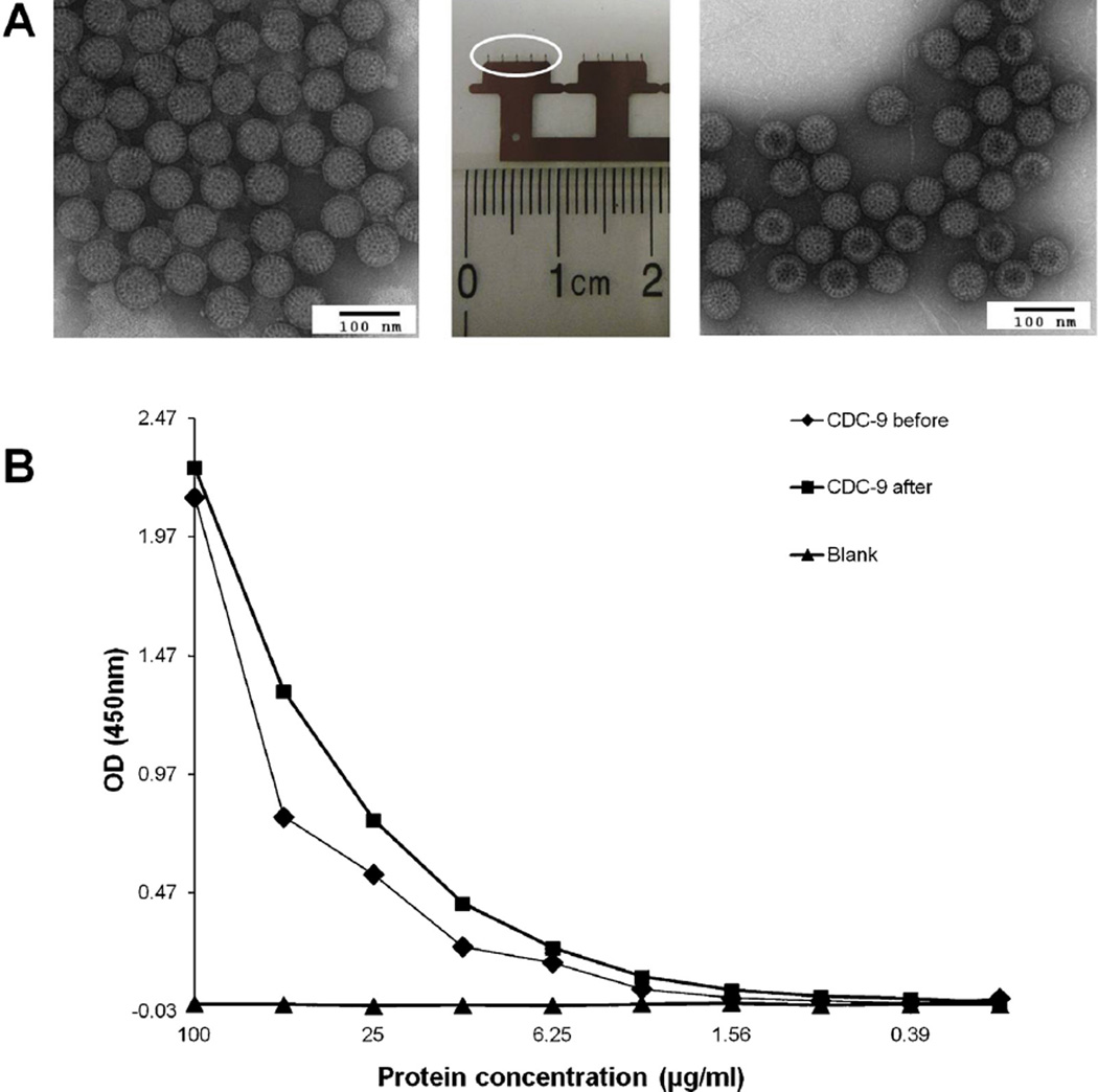
Stability and antigenicity of inactivated rotavirus particles coated on MN. A: Electron micrographs of inactivated CDC-9 IRV particles before coating (left) and after reconstitution from MN one day after coating (right). Triple-layered CDC-9 particles were stained with phosphotungstic acid and examined with an electron microscope. Bar = 100 nm. The central image shows two MN devices each with a row of five MN (circle shows a five-MN array). B: Levels of rotavirus antigen detected in original and reconstituted preparations by EIA. Similar levels of absorbance in original and reconstituted CDC9 preparations were observed. A blank was tested as a negative control.

