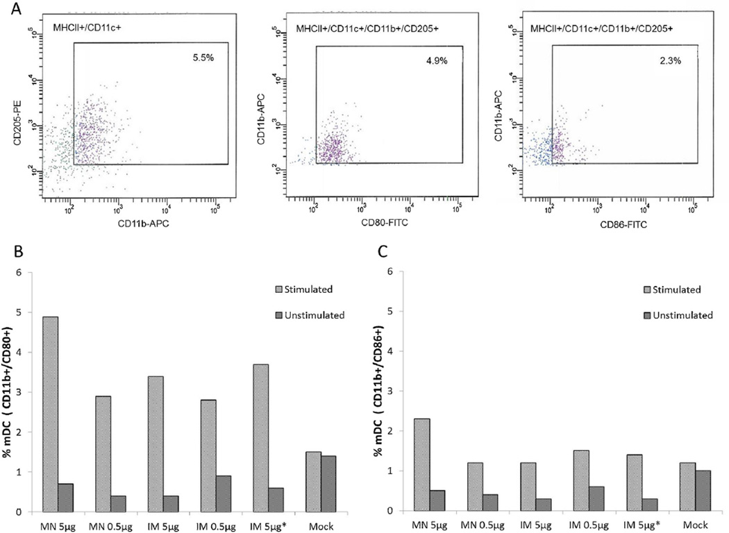
Fig. 2.
Phenotype and maturation of DCs in the spleens of mice that received IRV by MN and IM administration. Splenocytes were stimulated with rotavirus, purified for DCs, and examined for the expression of surface and co-stimulatory markers CD11c, CD80 and CD86 as described in the text. A: Representative FACS plots showing the phenotype and maturation expressing CD80 and CD86 of mDCs in the spleens of mice 28 days after receiving 5 µg of IRV using a MN patch, following in vitro stimulation with rotavirus. B and C: Proportions of activated mDCs with CD80 or CD86 expression in stimulated splenocytes of mice 28 days after receiving IRV by MN or IM administration. The character “*” indicates IRV reconstituted from coated MN.

