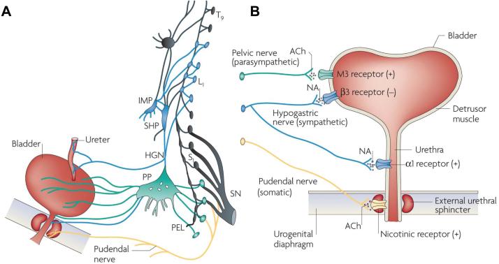
Figure 2.
Efferent pathways of the lower urinary tract. (A) Innervation of the female lower uri-nary tract. Sympathetic fibers (shown in blue) originate in the T11–L2 segments in the spinal cord and run through the inferior mesenteric ganglia (inferior mesenteric plexus [IMP]) and the hypogastric nerve [HGN]) or through the paravertebral chain to enter the pelvic nerves at the base of the bladder and urethra. Parasympathetic preganglionic fibers (shown in green) arise from the S2–S4 spinal segments and travel in sacral roots and pelvic nerves (PEL) to ganglia in the pelvic plexus (PP) and in the bladder wall. This is where the postganglionic nerves that supply parasympathetic innervation to the bladder arise. Somatic motor nerves (shown in yellow) that supply the striated muscles of the EUS arise from S2–S4 motor neurons and pass through the pudendal nerves. (B) Efferent pathways and neurotransmitter mechanisms that regulate the lower urinary tract. Parasympathetic postganglionic axons in the pelvic nerve release acetylcholine (ACh), which produces a bladder contraction by stimulating M3 muscarinic receptors in the bladder smooth muscle. Sympathetic postganglionic neurons release noradrenaline (NA), which activates β3 adrenergic receptors to relax bladder smooth muscle and activates α1 adrenergic receptors to contract urethral smooth muscle. Somatic axons in the pudendal nerve also release ACh, which produces a contraction of the external sphincter striated muscle by activating nicotinic cholinergic receptors. Parasympathetic postganglionic nerves also release ATP, which excites bladder smooth muscle, and nitric oxide, which relaxes urethral smooth muscle (not shown). L1, first lumbar root; S1, first sacral root; SHP, superior hypogastric plexus; SN, sciatic nerve; T9, ninth thoracic root. Reprinted with permission from (20): Fowler CJ, Griffiths D, de Groat WC. (2008) The neural control of micturition. Nat. Rev. Neurosci. 9:453–66.

