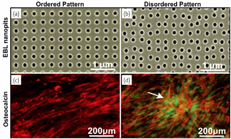
Fig. (10).
Different nanostructural patterns obtained using electron beam lithography (EBL), and the corresponding images of the expression of the bone-specific extracellular matrix protein osteopontin within differentiated mesenchymal stem cells. Note that the disordered pattern of the biomaterial constituents stimulates the protein expression more than the ordered pattern does. Reprinted with permission from Ref. [66].

