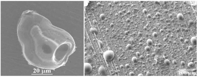
Fig. (7).
Another example of a drastic change in morphology following a minute modification of the experimental conditions [36]. Both morphologies shown were obtained after deswelling an identical, thixotropic mixture of cholesterol and carboxymethyl cellulose in an ultrasound field. The image on the left, however, corresponds to the particles prepared within a ten times smaller batch size compared with the morphology shown on the right. The same ultrasound intensity applied on a smaller volume in the former case breaks the material apart into smaller fragments, whereas it merely produces bubbles on a cholesteric surface in the system of a larger volume.

