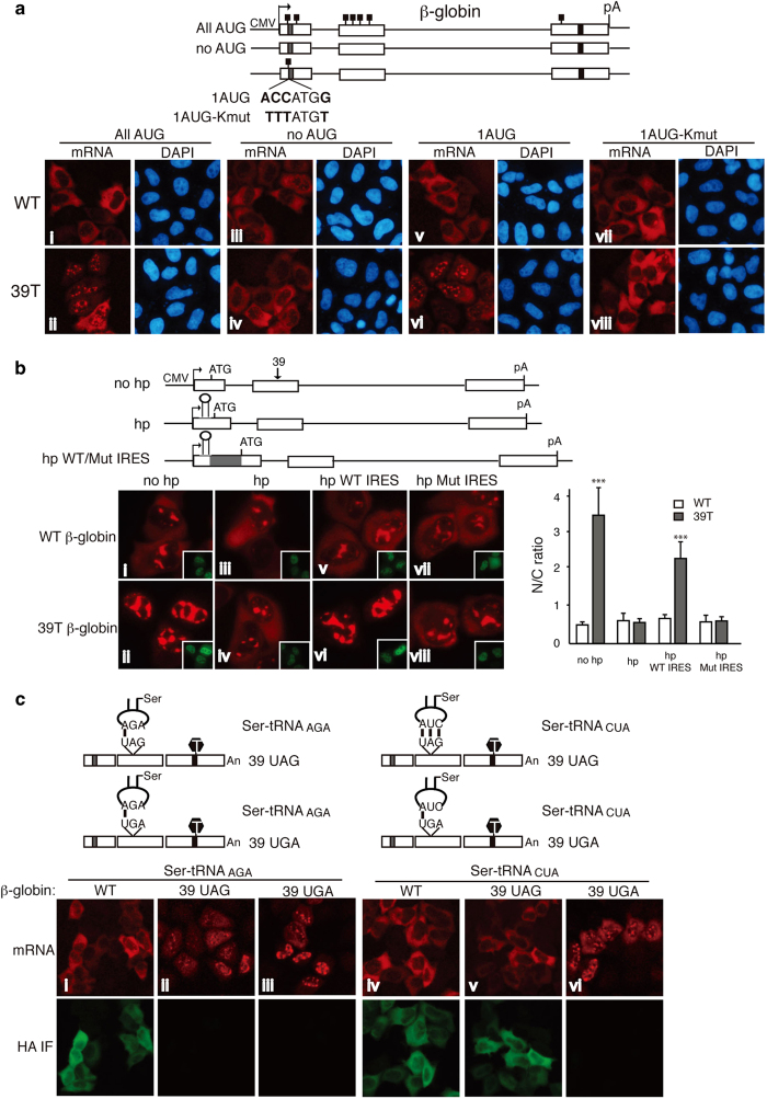
Figure 6.
Evidence that nuclear premature termination codon (PTC) recognition depends on the ribosome and tRNA. (a) Schematic of β-globin constructs is shown. The small black boxes indicate the ATGs. Equal amount of wild-type (WT) and mutant β-globin constructs were transfected into HeLa cells, and 12 h after transfection fluorescence in situ hybridization (FISH) was carried out. The percentages of cells with similar mRNA nucleocytoplasmic distribution patterns to the images shown were, from left to right, top, 84±4%, 83±3%, 90±3%, and 85±5%; bottom, 80±5%, 83±1%, 82±3%, and 88±3%. We have set the N/T ratio bigger than 1.2 as N-postive and smaller than 0.8 as C-positive. (b) Schematic of β-globin constructs. The gray box indicates the EMCV internal ribosomal entry sites (IRES). Positions of the start codon, the hairpin structure, and the PTC were marked. No hp, β-globin construct contains no hairpin; hp, β-globin construct contains a hairpin in the 5′ UTR; and hp WT/Mut IRES, β-globin construct contains a WT or mutant EMCV IRES downstream of the hairpin. β-Globin constructs were microinjected into the nuclei of HeLa cells, and α-amanitin was added 15 mins after microinjection. One hour after addition of α-amanitin, FISH was carried out to detect the mRNA. Inset pictures show the injection marker. The graph shows the average N/C ratios for β-globin mRNAs, and error bars indicate the standard errors among three independent experiments. Statistical analysis was performed as in Figure 1c. (c) Schematic of Ser-tRNA constructs whose anticodon either base pair or does not base pair with the PTC. Equal amount of WT and mutant Ser-tRNA constructs were co-transfected with WT or PTC+β-globin construct into HeLa cells, 12 h later, FISH and immunofluoresence with the HA antibody were carried out. The percentages of cells have similar mRNA nucleocytoplasmic distribution pattern to the images shown were, from left to right, 85±3%, 86±4%, 79±4%, 81±9%, 76±3%, and 77±2%. See also Supplementary Figures S5–S7.

