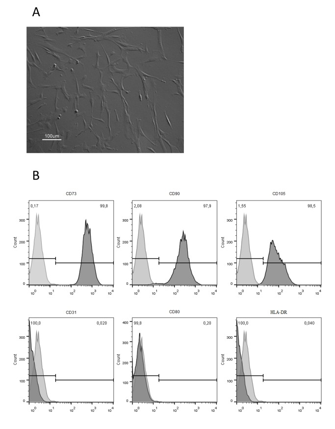
Fig 2. Characterization of bone marrow (BM)-derived mesenchymal stem/stromal cells (MSC) expanded on tissue culture flasks using DMEM culture medium supplemented with 10% FBS.
(A) Morphology of cultured cells assessed by optical microscopy. (B) Immunophenotype of cultured MSC assessed by flow cytometry according to Materials and Methods description. Percentage (%) displayed in the upper right corner of each histogram indicates the expression of each antigen.

