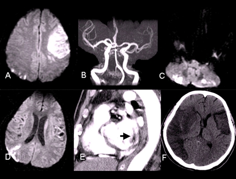
Figure 1.
(A) MRI of the secondary episode of stroke showed infarction of left frontal and right watershed area. (B) MRA of the secondary episode of stroke did not reveal significant focal stenosis. (C, D) MRI of the third episode of stroke disclosed punctuate stroke in several territories of the cerebral artery. (E) Multidetector computed tomography showed one filling defect in the posterior-lateral aspect of left atrium (arrow). (F) CT of the fourth episode of stroke showed infarct of complete right MCA territory with brain edema.

