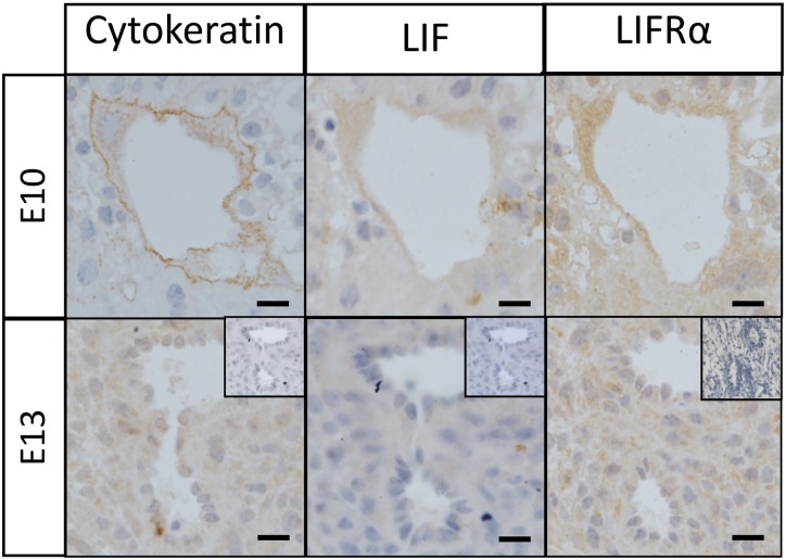
Fig 2. LIF and LIFRα co-localization with cytokeratin to detect invasive EVTs in wild-type mouse decidua.
Wild type (WT) mouse implantation sites were collected from n = 3 mice/time point and 2μm serial sections were immunostained for cytokeratin, LIF and LIFRα. Representative photomicrographs of mid-gestation (E10 and E13) implantation site sections are shown here. Both LIF and LIFRα co-localized with cytokeratin in maternal decidual vessels. Bars represent 20μm. Insets are negative controls.

