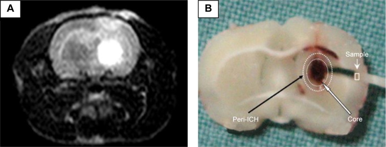
Figure 1.

Hematoma location and tissue collection.
Notes: (A) To confirm a striatal hematoma location, MRI was performed to track hemorrhage. On T2-weighted MRI, the hematoma appeared hyperintense in the striatum. (B) The fixed brain was dissected into slices through the coronal plane. In the center section, four 1- to 2-mm blocks of representative perihematomal brain tissue were obtained to assess mitochondrial structure. The circles denote the ICH core (inner circle) and its periphery (outer).
Abbreviations: MRI, magnetic resonance imaging; ICH, intracerebral hemorrhage.
