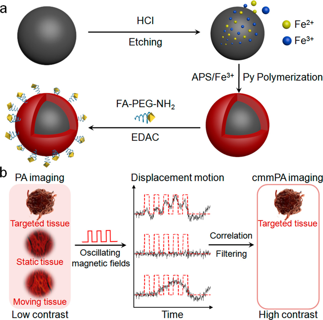
Figure 1.
Schematic of MNP–PPy core–shell nanoparticle fabrication and the mechanism of cyclic mmPA in imaging contrast enhancement. (a) Key steps involved in hybrid nanoparticle synthesis: Fe2+/Fe3+ generation though acid etching of monodisperse MNPs, PPy shell formation in the presence of Fe3+ and APS, and conjugation of FA–PEG–NH2 to nanoparticle surface. (b) Schematic of tumor detection via cyclic mmPA imaging. Contrast enhancement of tumor/background by suppressing both signals from static tissues and regions with random motions from moving tissues not related to the frequency of oscillating magnetic fields while identifying specific tumor signals from targeted tissues coherently responsive to cyclic magnetic motions.

