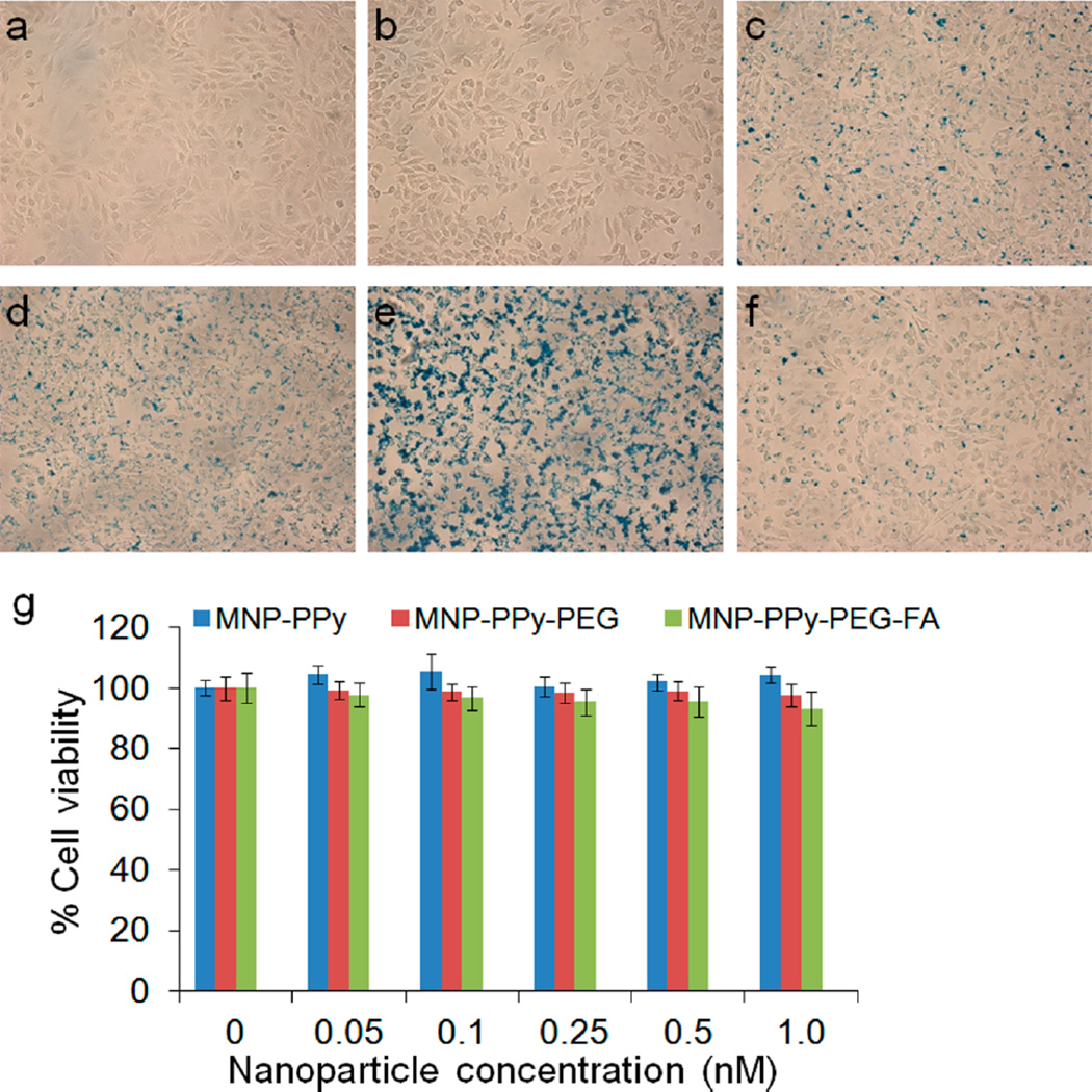
Figure 5.
Targeting specificity and cytotoxicity of hybrid nanoparticles. Bright field micrographs of HeLa cells (a) with no treatment and treated with (b) Prussian blue, (c) 0.5 nM MNP–PPy nanoparticles, (d) MNP–PPy–PEG nanoparticles, (e) MNP–PPy–PEG–FA nanoparticles, and (f) MNP–PPy–PEG–FA nanoparticles together with 1 mM free FA. (c–f) Stained with Prussian blue. (g) Dose-dependent cytotoxicity of MNP–PPy, MNP–PPy–PEG, and MNP–PPy–PEG–FA nanoparticles in HeLa cells plotted against the control groups with no treatment. In the concentration range probed between 0 and 1.0 nM, the three types of nanoparticles do not show significant cytotoxicity. Error bars represent standard deviations of three separate measurements.

