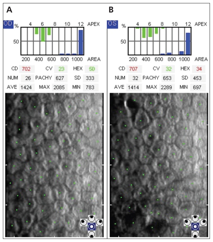
Figure 4:
Specular microscopy images showing the corneal endothelium, with each green dot corresponding to an individual cell. Decreased endothelial cell density is seen in the (A) right (702 cells/mm2) and (B) left eye (707 cells/mm2) at five-month follow-up. The normal cell density for this patient’s age group is between 2300 and 3100 cells/mm2.

