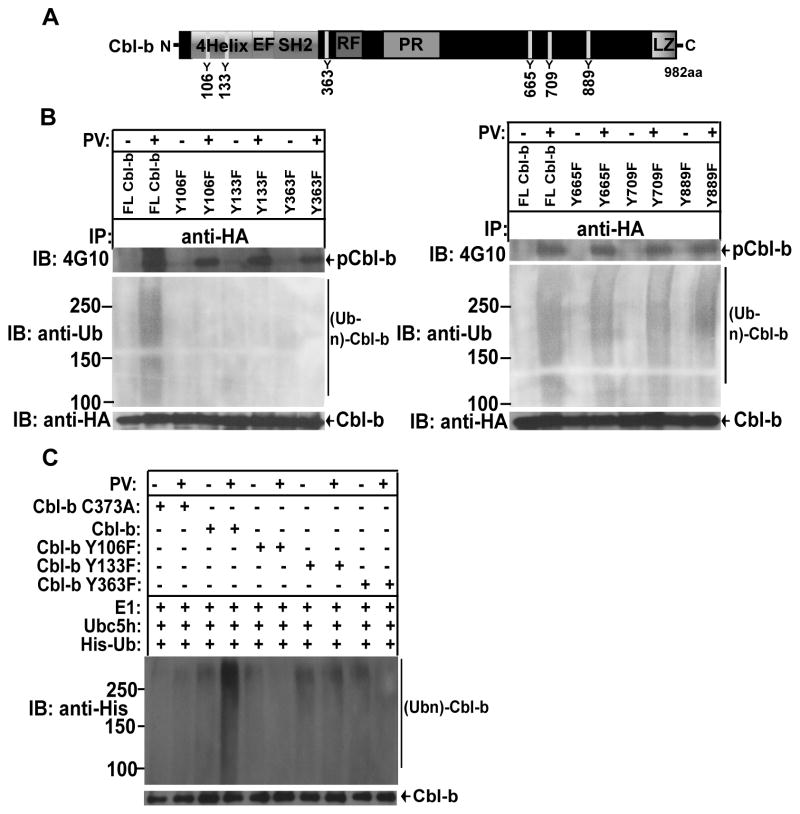
FIGURE 5. Cbl-b tyrosine phosphorylation at Y106, Y133, and Y363 is required for its activation.
(A) Schematic model of Cbl-b structure. (B) HEK293 cells transfected with HA-Cbl-b, or Cbl-b Y106F, Y133F, Y363F, Y665F, Y709F, and 889F, and treated with PV, and lysed in RIPA buffer. The cell lysates were immunoprecipitated with anti-HA, and blotted with 4G10. The membranes were stripped, and reprobed with anti-pTyr, anti-ubiquitin, and anti-HA. (C) Affinity-purified HA-Cbl-b, or Cbl-b Y106F, Y133F, Y363F, Y665F, Y709F, and 889F were incubated with E1, Ubc5, and His-tagged ubiquitin, and blotted with anti-His and anti-HA. Data are representative of two independent experiments.

