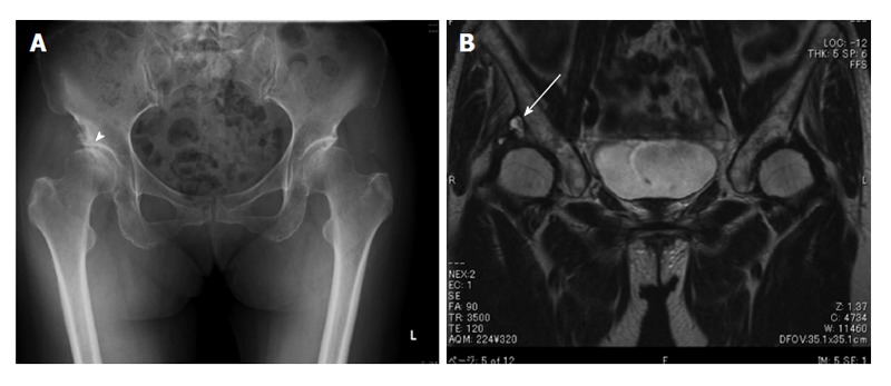
Figure 4.

A 70-year-old female with a cystic lesion associated with osteoarthritis. Plain radiograph of the lower pelvis reveals the secondary osteoarthritis of right hip due to acetabular dysplasia (arrowhead) (A). Coronal T2-weighted magnetic resonance image shows a cystic mass (arrow) located superiorly, connected to the right hip joint (B).
