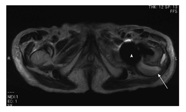
Figure 6.

A 93-year-old male with an infection after hemiarthroplasty for the left femoral neck fracture. Axial T2-weighted image reveals a cystic lesion with a fluid-fluid level (arrow) adjacent to the posterior aspect of the endoprosthesis (arrowhead).
