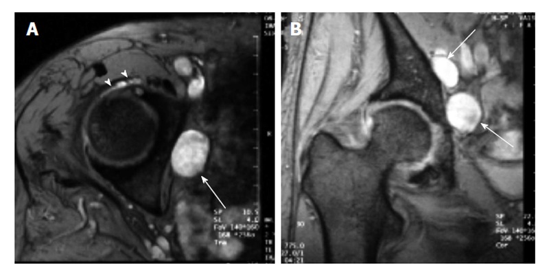
Figure 8.

A 75-year-old female with obturator neuropathy. Axial (A) and coronal (B) short tau inversion recovery magnetic resonance images show that the location of the cystic masses (arrows) is consistent with the site of obturator nerve (Ref: [26]). The stalk of the cyst was connected to the anterior joint capsule (arrowheads).
