
Figure 2.
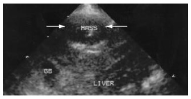
Image of VX2 tumor lesion under conventional second harmonic imaging. Arrow indicates the same VX2 tumor lesion at the anterior part of the right lobe. The echogenicity of the lesion and the around liver parenchyma seem unchanged at all.
Official websites use .gov
A
.gov website belongs to an official
government organization in the United States.
Secure .gov websites use HTTPS
A lock (
) or https:// means you've safely
connected to the .gov website. Share sensitive
information only on official, secure websites.
Image of VX2 tumor lesion under conventional second harmonic imaging. Arrow indicates the same VX2 tumor lesion at the anterior part of the right lobe. The echogenicity of the lesion and the around liver parenchyma seem unchanged at all.