
Figure 8.
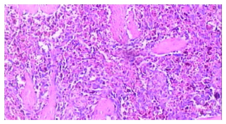
Microscopic examination of the thrombotic infarc-tion at the end of 4th week after RFA, the microthrombus dissolved, and extensive macrophages with hemosiderin depo-sition presented (HE. × 100).
Official websites use .gov
A
.gov website belongs to an official
government organization in the United States.
Secure .gov websites use HTTPS
A lock (
) or https:// means you've safely
connected to the .gov website. Share sensitive
information only on official, secure websites.
Microscopic examination of the thrombotic infarc-tion at the end of 4th week after RFA, the microthrombus dissolved, and extensive macrophages with hemosiderin depo-sition presented (HE. × 100).