
Figure 2.
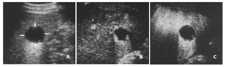
A 48-year-old man with metastasis of nasopharyngeal carcinoma. A. Intercostal section of precontrast conventional sonography exhibits a hypoechogenic lesion (arrows) with a diameter of 3.5 cm. B. Contrast-enhanced C-cube gray scale sonography at 24 s after injection of Levovist shows that peripheral enhancement appears at the same time with the liver parenchyma. C. Intranodular enhancement decreases at 107 s in the portal venous phase, earlier than that in the liver parenchyma.