
Figure 2.
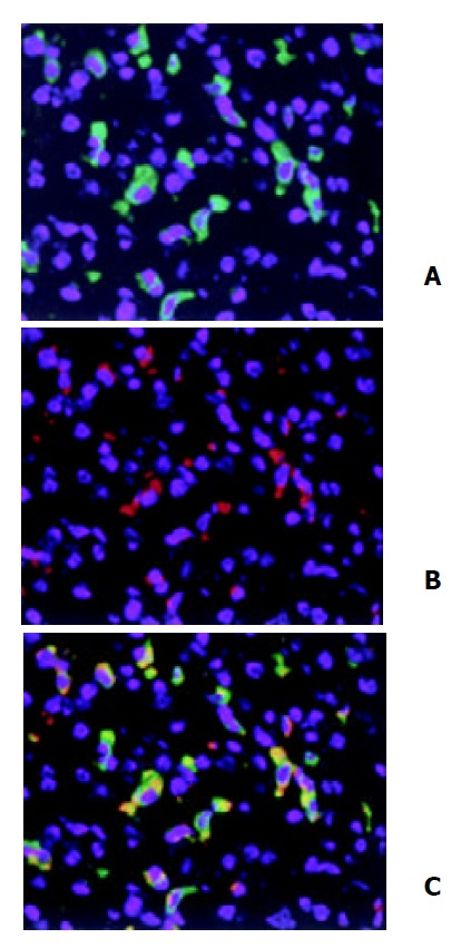
Double immunofluorescent staining of HO-1 local-ization in liver. After three doses of LPS treatment, HO-1 and CD68 in liver tissue were detected by double immunofluores-cent staining with polyclonal rabbit antibody against HO-1 followed by FITC-conjugated anti-rabbit IgG (green) and mono-clonal rat antibody against mouse CD68 followed by Cy3-con-jugated anti-rat IgG (red). Cell nuclei were counterstained with bis-benzimide (blue). A: HO-1; B: Macrophages (Kupffer cells); C: HO-1 + Macrophages. magnification × 400.