
Figure 2.
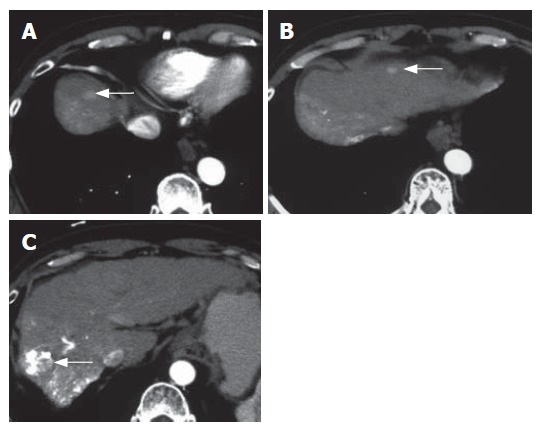
Contrast-enhanced CT at the time of the diagnosis of brain metastases shows multiple hypervascular HCCs (arrows) (A, B, C). Lipiodol retention in the main tumor is seen (C).
Official websites use .gov
A
.gov website belongs to an official
government organization in the United States.
Secure .gov websites use HTTPS
A lock (
) or https:// means you've safely
connected to the .gov website. Share sensitive
information only on official, secure websites.
Contrast-enhanced CT at the time of the diagnosis of brain metastases shows multiple hypervascular HCCs (arrows) (A, B, C). Lipiodol retention in the main tumor is seen (C).