
Figure 1.
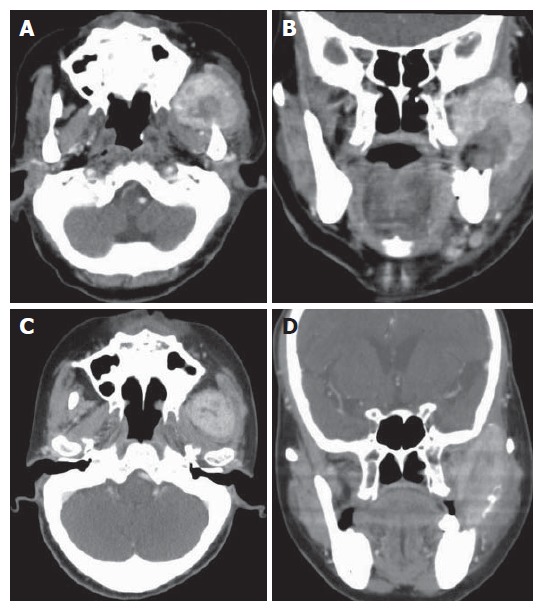
Axial and coronal CT scan of the neck with intravenous contrast showing a 6.2 cm х 5.0 cm, heterogeneously enhancing mass, which appears to be a left parapharyngeal mass involving the pterygoid muscle and temporal muscle (A and B); axial and coronal CT scan of the head and neck showing a 4.0 cm х 2.5 cm mass which shrunk after completion of radiotherapy (C and D).