
Figure 2.
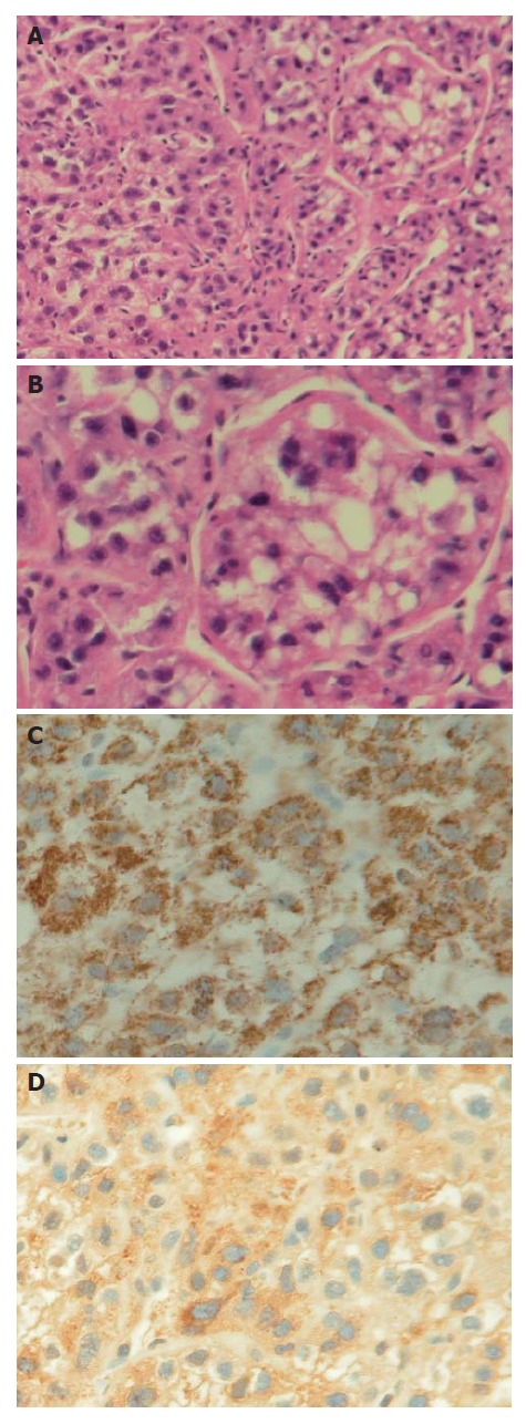
Histopathology of the metastatic lesion. A: The tumor is composed of polygonal cells featuring abundant clear to eosinophilic cytoplasm and microvesicular fatty change, and the tumor cells are arranged in broad trabeculae surrounded by sinusoid spaces which are characteristic of hepatocellular carcinoma (HE, х 100); B: Histology of the metastatic tumor (HE, x 200); C: The tumor cells showing immunoreactivity to Hep Par-1 with a cytoplasmic granular staining pattern (х 200); D: The tumor cells showing focal immunoreactivity to AFP (х 200).