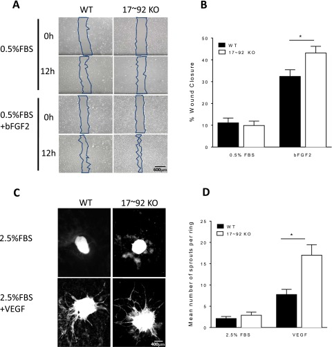
Fig. S4.
(A and B) Loss of miR 17∼92 KO in ECs augments recovery after wounding (A) and aortic sprouting (B) in vitro. Primary WT or 17∼92 KO ECs were starved, then wounded and treated with DMEM plus 0.5% FBS or 10 ng/mL bFGF2 for 12 h. The wound area was measured in captured images at 0 and 12 h, and migration was quantified. Data are duplicates from two experiments. (C) Aortic rings from WT and 17∼92VE-Cad mice were embedded in collagen type 1 and treated with +2.5% (vol/vol) FBS or VEGF 100 ng/mL for 6 d. At day 6, rings were fixed with PFA (4%) and stained with BS1 Lectin-FITC to delineate ECs. (D) Images of ring sprouting were obtained and quantified. Data are representative of five or six mice per group, and sprouts from eight or nine rings per mouse were quantified. Error bars represent mean ± SEM. *P < 0.05, one-way ANOVA with Bonferroni’s posttest. Data are representative of an experiment repeated three times and conducted in triplicate. *P < 0.05.

