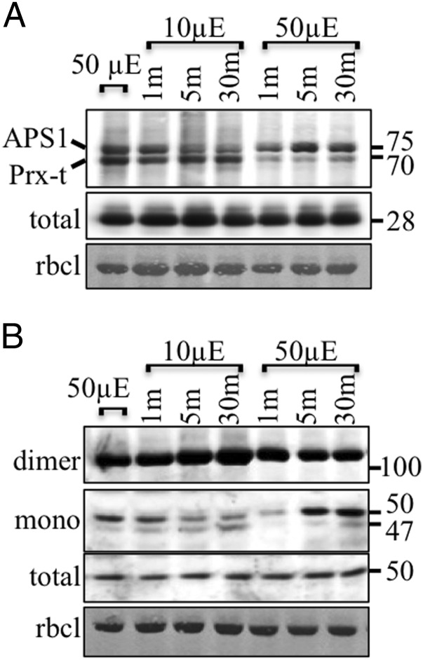
Fig. 3.
Immunoblot assay showing the ACHT4 intermolecular disulfide complexes (A) or APS1 redox state (B) in plants treated for 2 h with 50 μE·m−2·s−1 light intensity (50 µE) and after abrupt decreased (10 µE) followed by abrupt increased light intensity (50 µE). Equal protein loading was verified as in Fig. 1. The results shown are representative of three independent experiments.

