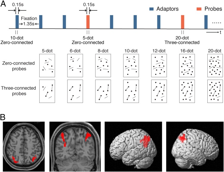
Fig. 5.
(A, Upper) Schematic description of the fMRI adaptation protocol and (A, Lower) illustration of probes containing 5–20 dots. Zero- and three-connected adaptors were actually the same as the zero- and three-connected probes containing 10 dots, respectively. Subjects were adapted to the zero- or three-connected 10-dot adaptors and tested with the zero- and three-connected probes containing variable numbers of dots. (B) Cluster of voxels sensitive to numerosity adaptation in the intraparietal sulcus from one representative subject shown on (Left) axial and coronal anatomical images and (Right) 3D-rendered lateral views. The voxels were defined by contrasting the activation of the zero-connected probes of 5 and 20 dots vs. the zero-connected probes of 10 dots.

