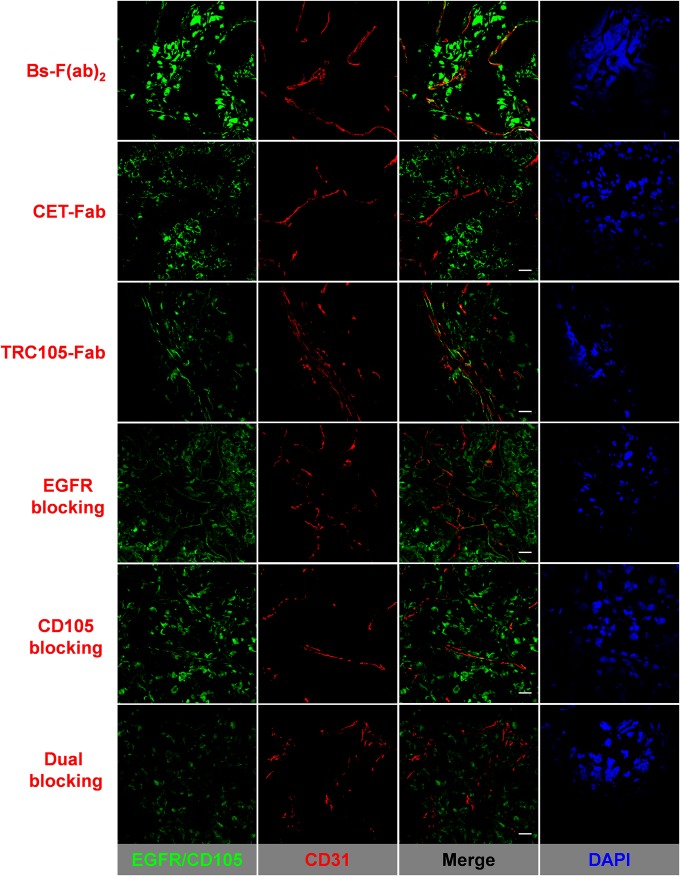
Fig. 4.
EGFR/CD105 immunofluorescence staining of resected U87MG tumors. FITC-labeled Bs-F(ab)2, CET-Fab, and TRC105-Fab were directly used for EGFR/CD105 staining (green). For blocking experiments, tissue slices were preincubated with 1 mg/mL of either cetuximab, TRC105, or a combination of both full mAbs. Rat anti-mouse CD31 antibody and Cy3-labeled donkey anti-rat IgG were used for CD31 staining (red). DAPI was used to stain cell nuclei. (Scale bar, 20 μm.)

