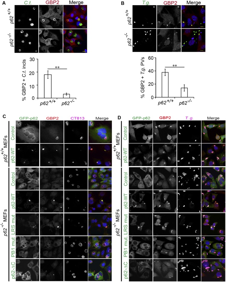
Fig. S8.
p62 promotes GBP2 recruitment to PVs. (A) WT p62+/+ and p62−/− MEFs were infected with GFP+ C. trachomatis, left untreated, or treated with IFNγ (100 U/mL) at 3 hpi. Cells were fixed at 20 hpi, stained with anti-GBP2 (red)/Hoechst (blue), and localization of endogenous GBP2 to inclusions was quantified. (B) IFNγ-primed (200 U/mL, overnight) and unprimed p62+/+ and p62−/− MEFs were infected with GFP+ T. gondii Pru. Localization of endogenous GBP2 to T. gondii PVs was monitored at 1 hpi. (C and D) Representative images of IFNγ-treated p62+/+ and p62−/− MEFs expressing the indicated p62 variants and infected with either C. trachomatis (C) or T. gondii Me49 (D). (Scale bar, 10 μm.) Frequency of GBP2 colocalizations with PVs was quantified, and data are shown in Fig. 6 C and D. **P < 0.005 (two-tailed unpaired t test). Data are representative of three independent experiments (mean ± SD; n = 3). (Scale bar, 10 μm.)

