Fig. 2.
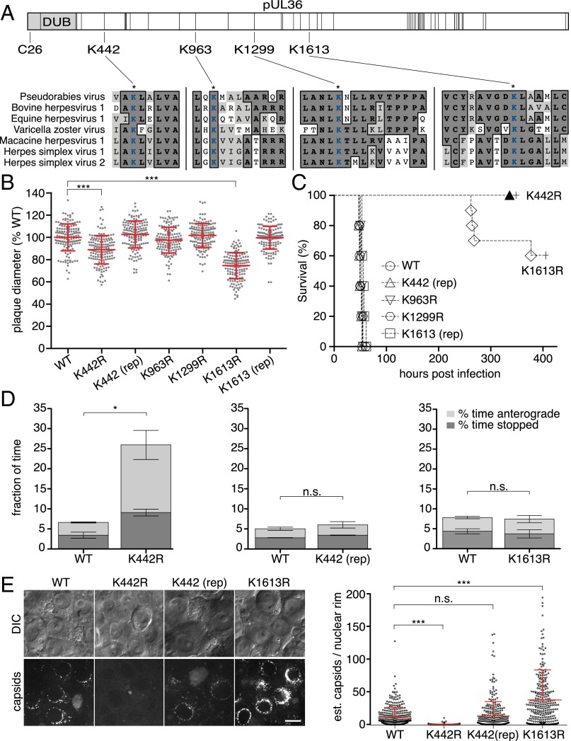
A conserved lysine in pUL36 is a virulence determinant that sustains retrograde axonal transport. (A) Illustration of PRV pUL36. Positions of lysine residues are noted with vertical bars, and the DUB is shown in light gray with the position of the active-site cysteine indicated. Four lysines conserved across seven neuroinvasive herpesviruses are shown with local sequence alignments (GenBank JF797219, AJ004801, AY665713, NC_001348, NC_004812, GU734771, and Z86099). (B) Spread of viruses in PK15 epithelial cells (n = 150 per sample; mean ± SD shown in red; ***P < 0.001 based on Tukey’s multiple-comparison test; rep, genetic repair). (C) Kaplan–Meier presentation of mouse survival following intranasal instillation of 106 plaque-forming units (PFU) of PRV (n = 10 for K442R and K1613R; n = 5 for all others). (D) Frequency of aberrant (nonretrograde) PRV axonal transport in cultured DRG sensory neurons 30–60 min postinfection (three independent experiments; n > 100 viral particles per sample; mean values ± SEM; *P < 0.05, two-tailed unpaired t test). (E) The number of fluorescent capsids at nuclear rims of cultured DRG (Left, representative images) was estimated (Right) at 3–4 hpi. (Scale bar: 10 μm.)

