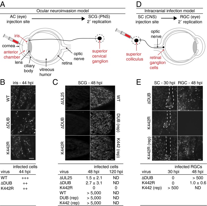
Fig. 4.
The herpes DUB and lysine substrate promote serial stages of neuroinvasion. (A and D) Two models of PRV retrograde axonal transport examined in this study (AC, anterior chamber; RGC, retinal ganglion cells; SC, superior colliculus; SCG, superior cervical ganglion). (A) Nerve endings are shielded by ocular tissue (iris) and not directly exposed to viral inoculum. (B and C) Representative images of the iris (B) and SCG (C) following intraocular injection of PRV strains encoding a mCherry-capsid reporter into the AC and retrograde transport to the SCG, with amount of infection based on fluorescent cell number indicated below. (D) Neurons and axons are directly exposed to viral inoculum. (E) Representative images and quantitation following intracranial injection into the SC and retrograde transmission to RGC of the contralateral eye. Microelectrode tracks are outlined in SC injection images to distinguish inoculum from viral spread. (Scale bars: 50 µm.)

