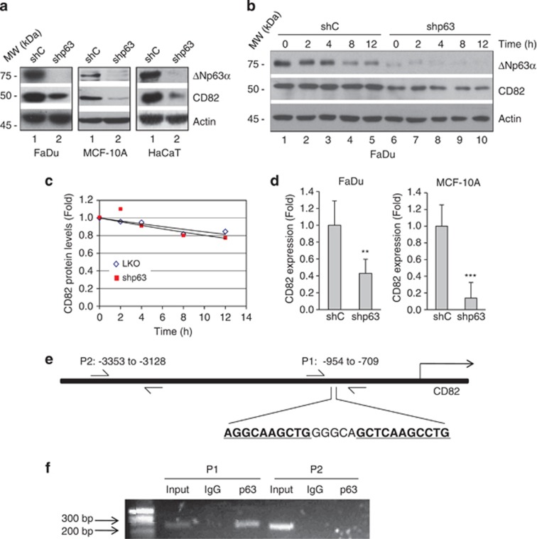
Figure 5.
CD82 is a transcriptional target of ΔNp63α. (a) FaDu, HaCaT and MCF-10A cells were infected with lentivirus expressing shRNA specific for pan-p63 (shp63) or a control shRNA (shC), and selected by puromycin resistance. Whole-cell lysates were subjected to western blot analysis, as shown. (b) FaDu cells expressing shp63 or shC were treated with 20 μg/ml cycloheximide for the indicated times. Whole-cell lysates were subjected to western blot analysis, as shown. (c) Quantitation of CD82 protein levels was performed using densitometry scanning. (d) Cells were subjected to Q-PCR analysis for CD82 expression. Results are presented as means and S.E. from three independent experiments performed in triplicate. **P<0.01; ***P<0.001. (e) Diagram of the CD82 gene and promoter locus showing a putative p63-binding element (P1: −954 to −709). An unrelated segment (P2: −3353 to −3128) was used as a negative control. Arrows indicate the position of the primers used for PCR amplification of the immunoprecipitated DNA. (f) Binding of p63 to this putative binding site was assessed in FaDu cells by chromatin immunoprecipitation using a specific p63 antibody (4A4) or a control mouse immunoglobulin G (IgG), followed by PCR amplification of both P1 and P2 sites

