Figure 1.
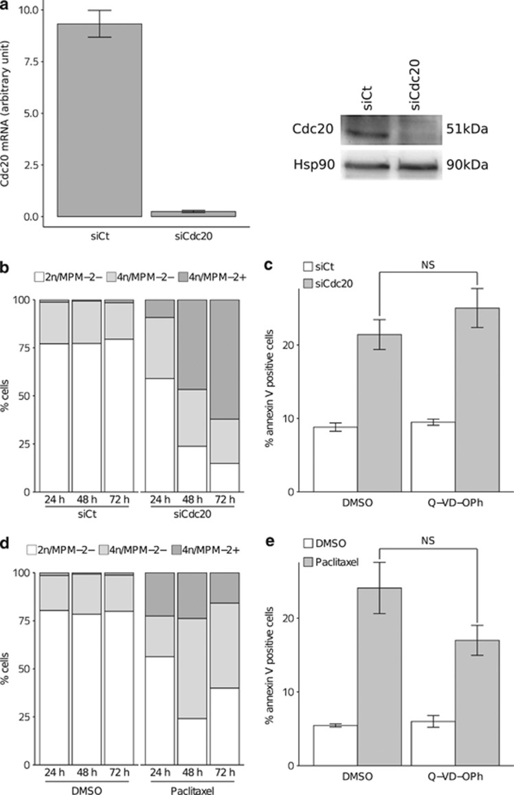
Cdc20 depletion triggered strong mitotic arrest followed by mitochondrial priming alteration, and caspase-independant cell death in breast cancer cells. (a) RT-qPCR and immunoblot analysis of Cdc20 expression levels after Cdc20 depletion by RNA interference in MDA-MB-231 cells. MDA-MB-231 cells were transfected with either a control siRNA (siCt) or a Cdc20 siRNA (siCdc20), and the Cdc20 mRNA (upper panel) or the protein (lower panel) levels were analysed using RT-qPCR and immunoblotting, respectively. (b) Cell cycle analysis of MDA-MB-231 cells under Cdc20 depletion. MDA-MB-231 cells were transfected with either a control siRNA (siCt) or a Cdc20 siRNA (siCdc20), and harvested at the indicated times. Cells were then stained with propidium iodide (PI) and MPM-2-directed antibody, and analysed by flow cytometry. 2n/MPM-2-negative, 4n/MPM-2-negative, 4n/MPM-2 positive cells corresponding to (G1/S), G2 or post-slippage cells, and mitotic (M) populations, respectively, are indicated. (c) Apoptosis analysis of MDA-MB-231 cells after Cdc20 depletion, and treatment or not with Q-VD-OPh, compared with control cells. MDA-MB-231 cells were transfected with either a control siRNA (siCt) or a Cdc20 siRNA (siCdc20), and treated with DMSO or the pan-caspase inhibitor Q-VD-OPh (10 μM). Cells were then stained with Annexin V, and analysed by flow cytometry. (d) Cell cycle analysis of MDA-MB-231 cells upon paclitaxel treatment. MDA-MB-231 cells were treated with 70 nM paclitaxel and harvested at the indicated times. Cells were then stained with PI and MPM-2-directed antibody, and analysed by flow cytometry. 2n/MPM-2-negative, 4n/MPM-2-negative, 4n/MPM-2-positive cells corresponding to (G1/S), G2 or post-slippage cells, and mitotic (M) populations, respectively, are indicated. (e) Apoptosis analysis of MDA-MB-231 cells after paclitaxel treatment in the presence of Q-VD-OPh or not, compared with control cells. MDA-MB-231 cells were pretreated with DMSO or the pan-caspase inhibitor Q-VD-OPh (10 μM) before paclitaxel treatment (70 nM). Cells were then stained with Annexin V, and analysed by flow cytometry

