Figure 5.
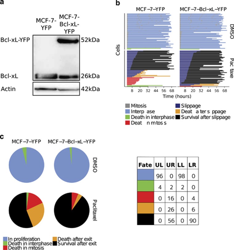
Bcl-xL overexpression protected cells from mitotic cell death. (a) Immunoblot analysis of Bcl-xL expression in MCF-7-YFP and MCF-7-Bcl-xL-YFP cells. (b) Fate profiles of MCF-7 cells expressing either YFP or Bcl-xL-YFP, and treated or not with paclitaxel. MCF-7-YFP and MCF-7-Bcl-xL-YFP cells were infected with H2B-RFP-coding lentivectors. Cells were then synchronized using a double thymidine block, treated with either DMSO or paclitaxel (70 nM), and finally analysed by time-lapse videomicrocopy during 72 h. Data for 50 cells per condition are presented. (c) End-point cell fates of MCF-7 cells expressing either YFP or Bcl-xL-YFP, and treated or paclitaxel. The proportion of each fate at the end point of experiments (72 h) derived from data presented in Figure 4b are shown in pie charts and % of cell fates in the associated table (UL=upper-left, UR=upper-right, LL=lower-left and LR=lower-right, corresponding panels in b)

