Figure 3.
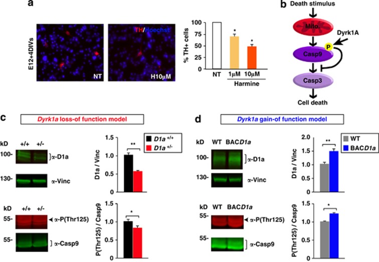
Effect of the DYRK1A inhibitor harmine on DA neuron survival and the levels of phosphorylated Casp9 in the developing ventral mesencephalon of Dyrk1a mutant mice. (a) Representative images of E12 mesencephalic cells after 4 days in culture (DIV) stained for TH (red), and the percentage of TH+ cells in cultures maintained in the presence or absence (NT) of harmine. (b) Scheme indicating the effect of Dyrk1a phosphorylation on the intrinsic cell death pathway. (c and d) Representative western blottings and their quantifications showing the levels of Dyrk1a (D1a) normalized to the amount of Vinculin (Vinc), and the proportion of Casp9 that is Thr125 phosphorylated (P(Thr125)) in total mesencephalic extracts from P2 Dyrk1a+/+ (D1a+/+) and Dyrk1a+/− (D1a+/−) mice (c), and from wild-type (WT) and mBACtgDyrk1a (BACD1a) mice (d). Mito, mitochondria. Histogram values are the mean±S.E.M. *P≤0.05; **P≤0.01 (n=3 in (a); n=4 in (c and d))

