
Figure 3.
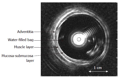
The cross-sectional ultrasound image of the distended distal esophagus allows identification of the esophageal layers, i.e. mucosa-submucosa, muscle and adventitia layers. The white round shadow in the centre is caused by the intraluminal ultrasound probe.